Carl Grisa finished his fight against cancer on Tuesday, Dec 13, 2011. Carl died, and the sarcoma died with him. Nobody won. He is in a better place now, without the cancer, BUT I WILL MISS HIM for the rest of my life.
Carl Grisa, 1992
Friday, Dec 16, 2011 we will have a service for Carl Grisa at Crossroads Presbyterian Church. There will be a closed casket visitation from 4:00 to 6:00 PM, Funeral Service from 6:00 to 7:00 PM and a Reception to follow. More details to follow.
Carl Grisa as I will remember him, 2007
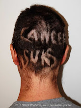
The cancer altered Carl's appearance. I hope the children will remember him as he was before cancer.
Figure A below is a MRI image of a sarcoma in Carl's neck as of Jan 5, 2011 with some added 'visual' information. In photo shop I added colors and labels, and drew a rough outline to help you orient the placement of the head, which hopefully helps you better understand the MRI.
In Figure A, the vertebrae (neck bones) are in green. The vertebrae part on the left in green (front of the neck) rings the spinal column and connects to the green part on the right (in the back of the neck).
The photo below shows a neck vertebrae bone as it would look if your were looking from the top of the head. You can see how the bone has a ring. The ring circles the spine.
The next visual labels different parts of the vertebrae.
In Figure A, the spinal column (in yellow) is protected by a fluid filled sack. Perhaps you can now see the bad news of Figure A.
The nasty sarcoma tumor (shown in red on Figure A) is impinging (almost touching) the spinal column. It is squishing the fluid filled spinal sack. Unfortunately, the sarcoma tumor has also eaten away and destroyed part of the bone in the back of the C2 vertebrae, and possibly others (C3?). Destroying parts of the bone of the vertebrae - Bad. Touching the spinal column (i.e. the spine) - Well, that would be way worse.
Below are images from a CT scan of Carl's head taken from the top of the head looking down. This CT scan is from January 5, 2011. CT scans show bone details clearly. You can see the vertebrae glowing in white. Instead of being shaped like the photo above, the white bone has been replaced on the right with ominous gray tumor material.
Below are three MRI images of the same cross section, again, from Jan 05, 2011. Above, CT scans, below MRI scans.
The three MRI images show different ways the MRI contrast can be adjusted to better see various types of tissues.
The final two images below are from a CT scan adjusted to show different structures. The views are a cross sections of Carl's neck from the back (image on the left), and a side view of the neck (on the right).
These images document why the sarcoma tumor in the neck had to be surgically removed. Carl had surgery to remove the tumor on January 24, 2011.
Click on any images to see it larger (and less blurry.)
When it comes to cancer care in 2011, in the United States, here in Wisconsin, the engineer and photographer in me is grateful and proud of the available digital diagnostic technology. The technology is improving exponentially.
Like the expansion of the Internet and the digital camera, diagnostic imaging is improving cancer detection, monitoring, and care. I don't like the fact that Carl has cancer, but if you have to get cancer, (and you can't delay getting cancer into the future), now is the time to get it over any other period in time, any other millennium, century or year. The 'technology', drugs, and care is improving. Teams of engineers, scientists, technicians and doctors have created the technology I am going attempt to show you below.
Don't let the fact that this technology is cool make you think I am in any way happy that my husband has cancer. The pep talk above does not make it any easier for me to use photo shop to try to document this tumor. I am doing this (originally) for my children; also my spouse, our relatives and friends; and others who may stumble across this post trying to understand cancer. I do not like spending time trying to make the images understandable. But I think I can use my time to help some people understand more about cancer, or Carl's cancer, where ever their interest might lie.
Below are some MRI images that show the tumor in Carl's neck as of Jan 5, 2011. MRI stands for Magnetic Resonance Imaging. See Wikipedia for technological information on MRI's. Basically, MRI's use a large machine, a lot of physics and a lot of engineering to take multiple cross section photos of a part of the body, in this case Carl's neck.
If you can't see how Figure 1 or Figure 2 are images of a view of a neck, see Figure 3 and 4 below for some added 'visual' information which may help you to be able to better understand the MRI's.
Figure 1 is a MRI of a sarcoma in Carl's neck as of Jan 5, 2011. This is a view as the doctor or technician can see it on their computer screen. On the computer, the doctor can manipulate settings like contrast.
Figure 2 is a MRI of a sarcoma in Carl's neck as of Jan 5, 2011 with different settings, as the doctor or technician can see it on their computer screen. Notice how the neck vertebrae (the bones) stand out differently then in Figure 1 above. In Figure 1 the vertebrae (bones) are gray, in Figure 2 the bones are dark and stand out more. The tumor is more visible in Figure 2 when the bones are dark.
Figure 3 is Figure 2 with some added 'visual' information. I took the Figure 2 above and in photoshop I added colors and labels, and drew a childish outline to help you orient the placement of the head, which hopefully helps you better understand the MRI. Figure 3 is NOT what the doctors see on their computer screens.
Figure 4 is Figure 2 with some added 'visual' information. It is my first attempt at showing what parts are what. I took the Figure 2 above and in photo shop I added labels, and drew a childish outline to help you orient the placement of the head. Figure 4 is NOT what the doctors see on their computer screens.
MRI's are good for showing tissues, and differences in tissues. Carl also gets CAT scans. CAT scans are good at showing bone and structures like veins. I hope to show you CAT scans on a different future post. Carl also gets some X-Rays, which are good for showing bones, although differently then in a CAT scan. The combination of MRI's and CAT scans give doctors knowledge (and a visual picture) of things inside the body that used to be able to only be seen once a surgeon cut the body open. Now, the doctors and surgeons learn much before any cuts are made.
My children find these photos interesting for several seconds, and then at some point the information creeps them out and they need to walk away. It is not just a screen shot, it is a tumor that once was in their dad. (Surgery removed almost all of the tumor in Carl's neck on Monday Jan 24, 2011.) I am telling you about my 11-16 year old children's responses so that you may be aware of and accept your own feelings. I find it easiest to view these images when I detach them from being my husband's neck images. (It is easier to view them 'clinically'.)
I waited months to post these images. Maybe you will understand why. Whatever.
The next blog post will talk about Carl's MRI more specifically.
Click on any images to see it larger (and less blurry.)
We last left off December 22, 2009, the happy day where Carl got his port placed. Many of these fun new terms (ex. port) will be defined in later posts.
2009 Dec 23, Wednesday:
We woke up calm and relaxed, a plan (and port) in place. We got the children off to school.
Carl was showered and dressed, ready to go to work for a nearly full day.
We get a phone call. Would we like to meet with the Radiologist? (by the way, TODAY?)
11:00 AM Meeting with the radiologist.
The thing in your neck concerns us... (what thing?) Remember the PET scan on December 18? The first Scan including the head? Guess what...
1:00 PM Hi tech custom device to immobilize Carl's head and neck for radiation created (for photo see What Radiation looks like.)
CT simulation of radiation to neck area done.
I left Carl at Froedtert to collect the children from the last day of school for the year 2009.
4:00 PM Carl got Radiation #1 (of 10) and was driven home by friend. (see What Radiation looks like)
The treat in treatments are starting, before Christmas even.
4:50 PM I got a call from an anguished Anthony. He was hurt at wrestling practice.
5:15 PM Carl home from Radiation #1.
5:40-7:45 PM Mom and Anthony at the urgent care facility, X-ray shows a totally broken collar bone with the two pieces of bone not even close to touching.
8:30 PM Pain medication prescription filled for Anthony and Carl.
Lots of Christmas snow coming down.
Children at home with Carl put up the Christmas ornaments on tree (front side only).
2009 Dec 24, Thursday:
The children slept in at home - no school.
8:00 AM Carl got Radiation #2 (of 10) at Froedtert Hospital in Milwaukee, Wisconsin.
Carl got a neck brace. (You have to be kidding, right?) After Carl indicated he was unlikely to wear the neck brace the nurse told him that one of his vertebrae had some bone that was partially missing... You don't want to be paralyzed, do you? (answer, nope, and Carl doesn't want a sarcoma either.)
New song invented. "It's beginning to look a lot like cancer, Every-where they look..." [Sing to the familiar Christmas tune.]
We went home and collected the children. They enjoyed the non-stop screen time while we were gone very much.
Christmas Eve Party with family on my side of the family. Carl moved his head carefully and didn't wear the neck brace. He figured he would risk paralysis for one more day.
I went home and Carl and I went to bed. Carl slept with the neck brace on. I sighed, then got up and wrapped the presents I had and put them under the tree.
2009 Dec 25, Friday:
Santa left precisely 5 wrapped presents under our glowing (1200 lights!) tree. This does not compare favorably to the bounty from previous years. The children did NOT let us sleep in, and one present WAS open before all the family was assembled.
The children still had a good Christmas anyway (It's not like we don't have any toys, after all), and a Shopping Spree Day for Saturday was discussed.
Christmas Party with family on Carl's side of the family. Carl wore the neck brace. "Let the Freak show begin" Carl said as he walked in with his neck brace and cane. OK, this is not a great thing for Carl to say, and I probably should leave it out, but it's what he said, it's what he felt, and it's what I report here. Carl thinks he will blend more once his hair falls out.
2009 Dec 26, Saturday:
Shopping Spree day with family and Uncle Paul. We got the children pretty much whatever they wanted [but alas, no XBOX 360, Anthony's dearest wish. Had the progress reports of multiple children shown more (some?) progress...]
Lots of snow falling fast does not dissuade shoppers.
Parking was atrocious at the stores and malls. Even handicapped parking was very full (Carl got a temporary disabled parking sticker with the cane). Carl thinks they must be giving temporary disabled parking stickers out to anyone who asks. (OK, lots of the humor going around is kind of dark at the moment...)
2009 Dec 27, Sunday:
11:00 AM Church service. (Carl, with neck brace and cane attracted some welcome attention - I love my church family! Thank you all!)
2009 Monday Dec 28, to Thursday Dec 31... :
7:30 Carl got Radiation
8:00 Carl got Chemotherapy
Carl did other tests, labs, appointments...
Christmas and December 2009 will be remembered like no other... Each day of Dec 28, Dec 29, Dec 30, and Dec 31 were their own mini adventures... But I am now tired and will post this post, and I will have covered 2009. Sort of.
Happy New Year to all. (This I mean sincerely).
This is the first year in more then 15 years that I have not had my Christmas cards made and sent out before the New Year started.Something else to do in January 2010.I must make the cards, and I must get the photo, before Carl's hair falls out. After all, I scrapbook my Christmas cards into my Christmas Album. For that reason alone, if none other, I intend to create the Christmas cards, and send them out! [Note from May, 2010: I never sent out Christmas cards for 2009]
This blog is about Carl Grisa's journey / challenge / experience / fight with cancer; written mostly by his wife to document and share with friends, relatives, and other interested parties Carl Grisa's journey.