Figure A below is a MRI image of a sarcoma in Carl's neck as of Jan 5, 2011 with some added 'visual' information. In photo shop I added colors and labels, and drew a rough outline to help you orient the placement of the head, which hopefully helps you better understand the MRI.
 In Figure A, the vertebrae (neck bones) are in green. The vertebrae part on the left in green (front of the neck) rings the spinal column and connects to the green part on the right (in the back of the neck).
The photo below shows a neck vertebrae bone as it would look if your were looking from the top of the head. You can see how the bone has a ring. The ring circles the spine.
In Figure A, the vertebrae (neck bones) are in green. The vertebrae part on the left in green (front of the neck) rings the spinal column and connects to the green part on the right (in the back of the neck).
The photo below shows a neck vertebrae bone as it would look if your were looking from the top of the head. You can see how the bone has a ring. The ring circles the spine.
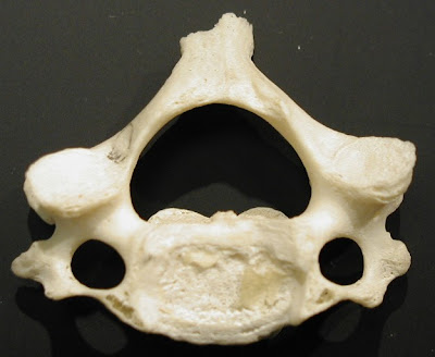
The next visual labels different parts of the vertebrae.
 In Figure A, the spinal column (in yellow) is protected by a fluid filled sack. Perhaps you can now see the bad news of Figure A.
The nasty sarcoma tumor (shown in red on Figure A) is impinging (almost touching) the spinal column. It is squishing the fluid filled spinal sack. Unfortunately, the sarcoma tumor has also eaten away and destroyed part of the bone in the back of the C2 vertebrae, and possibly others (C3?). Destroying parts of the bone of the vertebrae - Bad. Touching the spinal column (i.e. the spine) - Well, that would be way worse.
Below are images from a CT scan of Carl's head taken from the top of the head looking down. This CT scan is from January 5, 2011. CT scans show bone details clearly. You can see the vertebrae glowing in white. Instead of being shaped like the photo above, the white bone has been replaced on the right with ominous gray tumor material.
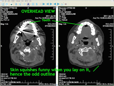
Below are three MRI images of the same cross section, again, from Jan 05, 2011. Above, CT scans, below MRI scans.


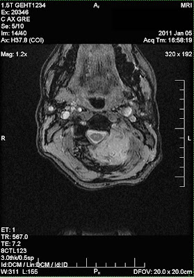
The three MRI images show different ways the MRI contrast can be adjusted to better see various types of tissues.
The final two images below are from a CT scan adjusted to show different structures. The views are a cross sections of Carl's neck from the back (image on the left), and a side view of the neck (on the right).
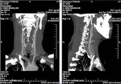 These images document why the sarcoma tumor in the neck had to be surgically removed. Carl had surgery to remove the tumor on January 24, 2011.
Click on any images to see it larger (and less blurry.)
When it comes to cancer care in 2011, in the United States, here in Wisconsin, the engineer and photographer in me is grateful and proud of the available digital diagnostic technology. The technology is improving exponentially.
Like the expansion of the Internet and the digital camera, diagnostic imaging is improving cancer detection, monitoring, and care. I don't like the fact that Carl has cancer, but if you have to get cancer, (and you can't delay getting cancer into the future), now is the time to get it over any other period in time, any other millennium, century or year. The 'technology', drugs, and care is improving. Teams of engineers, scientists, technicians and doctors have created the technology I am going attempt to show you below.
Don't let the fact that this technology is cool make you think I am in any way happy that my husband has cancer. The pep talk above does not make it any easier for me to use photo shop to try to document this tumor. I am doing this (originally) for my children; also my spouse, our relatives and friends; and others who may stumble across this post trying to understand cancer. I do not like spending time trying to make the images understandable. But I think I can use my time to help some people understand more about cancer, or Carl's cancer, where ever their interest might lie.
Below are some MRI images that show the tumor in Carl's neck as of Jan 5, 2011. MRI stands for Magnetic Resonance Imaging. See Wikipedia for technological information on MRI's. Basically, MRI's use a large machine, a lot of physics and a lot of engineering to take multiple cross section photos of a part of the body, in this case Carl's neck.
If you can't see how Figure 1 or Figure 2 are images of a view of a neck, see Figure 3 and 4 below for some added 'visual' information which may help you to be able to better understand the MRI's.
Figure 1 is a MRI of a sarcoma in Carl's neck as of Jan 5, 2011. This is a view as the doctor or technician can see it on their computer screen. On the computer, the doctor can manipulate settings like contrast.
 Figure 2 is a MRI of a sarcoma in Carl's neck as of Jan 5, 2011 with different settings, as the doctor or technician can see it on their computer screen. Notice how the neck vertebrae (the bones) stand out differently then in Figure 1 above. In Figure 1 the vertebrae (bones) are gray, in Figure 2 the bones are dark and stand out more. The tumor is more visible in Figure 2 when the bones are dark.
 Figure 3 is Figure 2 with some added 'visual' information. I took the Figure 2 above and in photoshop I added colors and labels, and drew a childish outline to help you orient the placement of the head, which hopefully helps you better understand the MRI. Figure 3 is NOT what the doctors see on their computer screens.
 Figure 4 is Figure 2 with some added 'visual' information. It is my first attempt at showing what parts are what. I took the Figure 2 above and in photo shop I added labels, and drew a childish outline to help you orient the placement of the head. Figure 4 is NOT what the doctors see on their computer screens.
 MRI's are good for showing tissues, and differences in tissues. Carl also gets CAT scans. CAT scans are good at showing bone and structures like veins. I hope to show you CAT scans on a different future post. Carl also gets some X-Rays, which are good for showing bones, although differently then in a CAT scan. The combination of MRI's and CAT scans give doctors knowledge (and a visual picture) of things inside the body that used to be able to only be seen once a surgeon cut the body open. Now, the doctors and surgeons learn much before any cuts are made.
My children find these photos interesting for several seconds, and then at some point the information creeps them out and they need to walk away. It is not just a screen shot, it is a tumor that once was in their dad. (Surgery removed almost all of the tumor in Carl's neck on Monday Jan 24, 2011.) I am telling you about my 11-16 year old children's responses so that you may be aware of and accept your own feelings. I find it easiest to view these images when I detach them from being my husband's neck images. (It is easier to view them 'clinically'.)
I waited months to post these images. Maybe you will understand why. Whatever.
The next blog post will talk about Carl's MRI more specifically.
MRI's are good for showing tissues, and differences in tissues. Carl also gets CAT scans. CAT scans are good at showing bone and structures like veins. I hope to show you CAT scans on a different future post. Carl also gets some X-Rays, which are good for showing bones, although differently then in a CAT scan. The combination of MRI's and CAT scans give doctors knowledge (and a visual picture) of things inside the body that used to be able to only be seen once a surgeon cut the body open. Now, the doctors and surgeons learn much before any cuts are made.
My children find these photos interesting for several seconds, and then at some point the information creeps them out and they need to walk away. It is not just a screen shot, it is a tumor that once was in their dad. (Surgery removed almost all of the tumor in Carl's neck on Monday Jan 24, 2011.) I am telling you about my 11-16 year old children's responses so that you may be aware of and accept your own feelings. I find it easiest to view these images when I detach them from being my husband's neck images. (It is easier to view them 'clinically'.)
I waited months to post these images. Maybe you will understand why. Whatever.
The next blog post will talk about Carl's MRI more specifically.
Click on any images to see it larger (and less blurry.)