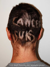
The children are back in school, and Carl is back to getting chemotherapy.
The children started school September 1, 2011, and have had four days of school so far. Tom is a High School Junior (11th grade), Anthony is a High School Sophmore (10th grade), Nathan is in 8th grade, and Miranda is in 7th grade.
Carl started an overnight 24 hour infusion on Wednesday, September 7, 2011, and that chemotherapy infusion is ending today, Thursday, September 8, 2011. It is a new chemotherapy drug for Carl - an experimental drug with some weird name. (I'll find out the name later)
I would love lots of prayers that this new chemotherapy drug works wonders - the stupid cancer cells have been growing again. Please put Carl on your church's prayers list as well.
I guess I shouldn't be surprised that some cells are growing, since Carl has had only one other chemotherapy course in 2011, but I admit that the new cells are discouraging.
By the way, Carl started out this chemotherapy course weighing 136 lbs.
9978
Showing posts with label neck tumor. Show all posts
Showing posts with label neck tumor. Show all posts
Thursday, September 8, 2011


Wednesday, June 15, 2011
Neck Surgery No. 3
Carl was wheeled in to surgery at 8:30 AM. at Froedtert Hospital, and there now as I am typing this.
I hope to update my blog with details later this week.
Monday, March 28, 2011
Neck MRI and CT scan images
Figure A below is a MRI image of a sarcoma in Carl's neck as of Jan 5, 2011 with some added 'visual' information. In photo shop I added colors and labels, and drew a rough outline to help you orient the placement of the head, which hopefully helps you better understand the MRI.

In Figure A, the vertebrae (neck bones) are in green. The vertebrae part on the left in green (front of the neck) rings the spinal column and connects to the green part on the right (in the back of the neck).
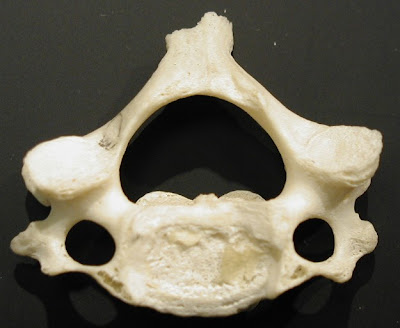
The photo below shows a neck vertebrae bone as it would look if your were looking from the top of the head. You can see how the bone has a ring. The ring circles the spine.
The next visual labels different parts of the vertebrae.

In Figure A, the spinal column (in yellow) is protected by a fluid filled sack. Perhaps you can now see the bad news of Figure A.
The nasty sarcoma tumor (shown in red on Figure A) is impinging (almost touching) the spinal column. It is squishing the fluid filled spinal sack. Unfortunately, the sarcoma tumor has also eaten away and destroyed part of the bone in the back of the C2 vertebrae, and possibly others (C3?). Destroying parts of the bone of the vertebrae - Bad. Touching the spinal column (i.e. the spine) - Well, that would be way worse.
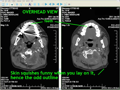
Below are images from a CT scan of Carl's head taken from the top of the head looking down. This CT scan is from January 5, 2011. CT scans show bone details clearly. You can see the vertebrae glowing in white. Instead of being shaped like the photo above, the white bone has been replaced on the right with ominous gray tumor material.
Below are three MRI images of the same cross section, again, from Jan 05, 2011. Above, CT scans, below MRI scans.


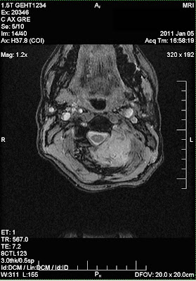
The three MRI images show different ways the MRI contrast can be adjusted to better see various types of tissues.
The final two images below are from a CT scan adjusted to show different structures. The views are a cross sections of Carl's neck from the back (image on the left), and a side view of the neck (on the right).
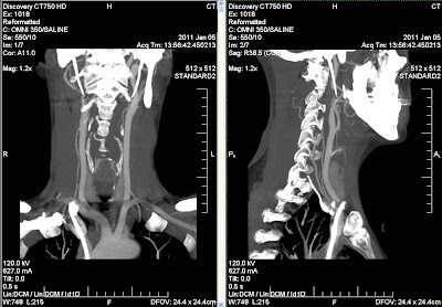
These images document why the sarcoma tumor in the neck had to be surgically removed. Carl had surgery to remove the tumor on January 24, 2011.
Click on any images to see it larger (and less blurry.)

In Figure A, the vertebrae (neck bones) are in green. The vertebrae part on the left in green (front of the neck) rings the spinal column and connects to the green part on the right (in the back of the neck).
The photo below shows a neck vertebrae bone as it would look if your were looking from the top of the head. You can see how the bone has a ring. The ring circles the spine.

The next visual labels different parts of the vertebrae.

In Figure A, the spinal column (in yellow) is protected by a fluid filled sack. Perhaps you can now see the bad news of Figure A.
The nasty sarcoma tumor (shown in red on Figure A) is impinging (almost touching) the spinal column. It is squishing the fluid filled spinal sack. Unfortunately, the sarcoma tumor has also eaten away and destroyed part of the bone in the back of the C2 vertebrae, and possibly others (C3?). Destroying parts of the bone of the vertebrae - Bad. Touching the spinal column (i.e. the spine) - Well, that would be way worse.
Below are images from a CT scan of Carl's head taken from the top of the head looking down. This CT scan is from January 5, 2011. CT scans show bone details clearly. You can see the vertebrae glowing in white. Instead of being shaped like the photo above, the white bone has been replaced on the right with ominous gray tumor material.

Below are three MRI images of the same cross section, again, from Jan 05, 2011. Above, CT scans, below MRI scans.


The three MRI images show different ways the MRI contrast can be adjusted to better see various types of tissues.
The final two images below are from a CT scan adjusted to show different structures. The views are a cross sections of Carl's neck from the back (image on the left), and a side view of the neck (on the right).

These images document why the sarcoma tumor in the neck had to be surgically removed. Carl had surgery to remove the tumor on January 24, 2011.
Click on any images to see it larger (and less blurry.)
Subscribe to:
Posts (Atom)