In Figure A, the vertebrae (neck bones) are in green. The vertebrae part on the left in green (front of the neck) rings the spinal column and connects to the green part on the right (in the back of the neck).
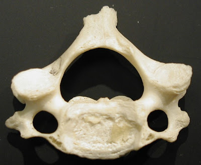
The photo below shows a neck vertebrae bone as it would look if your were looking from the top of the head. You can see how the bone has a ring. The ring circles the spine.

The next visual labels different parts of the vertebrae.

In Figure A, the spinal column (in yellow) is protected by a fluid filled sack. Perhaps you can now see the bad news of Figure A.
The nasty sarcoma tumor (shown in red on Figure A) is impinging (almost touching) the spinal column. It is squishing the fluid filled spinal sack. Unfortunately, the sarcoma tumor has also eaten away and destroyed part of the bone in the back of the C2 vertebrae, and possibly others (C3?). Destroying parts of the bone of the vertebrae - Bad. Touching the spinal column (i.e. the spine) - Well, that would be way worse.
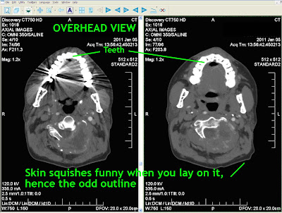
Below are images from a CT scan of Carl's head taken from the top of the head looking down. This CT scan is from January 5, 2011. CT scans show bone details clearly. You can see the vertebrae glowing in white. Instead of being shaped like the photo above, the white bone has been replaced on the right with ominous gray tumor material.

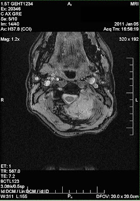
Below are three MRI images of the same cross section, again, from Jan 05, 2011. Above, CT scans, below MRI scans.


The three MRI images show different ways the MRI contrast can be adjusted to better see various types of tissues.
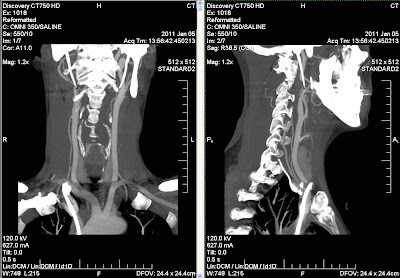
The final two images below are from a CT scan adjusted to show different structures. The views are a cross sections of Carl's neck from the back (image on the left), and a side view of the neck (on the right).

These images document why the sarcoma tumor in the neck had to be surgically removed. Carl had surgery to remove the tumor on January 24, 2011.
Click on any images to see it larger (and less blurry.)


2 comments:
Can you tell me the name of the corkscrew shaped gland that begins at the clavical and it's purpose? Please e-mail me karynglasser@yahoo.com
It is not my first time to go to see this website, i
am browsing this web page dailly and take pleasant information from here
all the time.
Here is my web page http://safedietplans.com
Also see my web page > diets that work
Post a Comment